Diagnostic Images with Enhanced Tube and Line Visibility in Mobile CXR
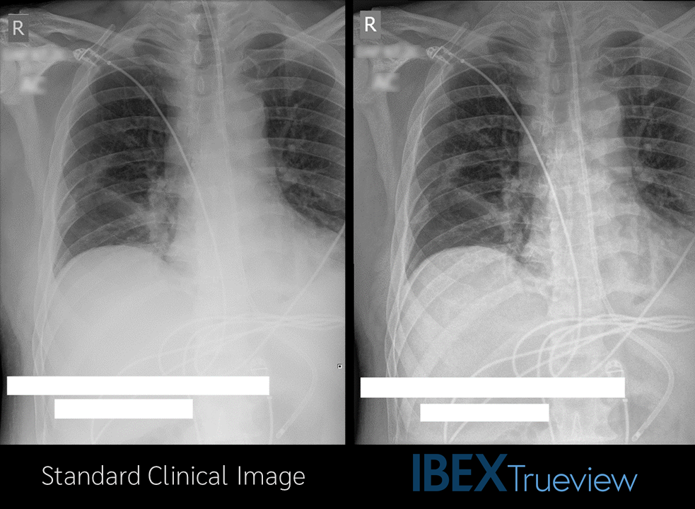
Tube and line visualisation is a significant challenge in chest X-rays collected in the ITU setting. Anti-scatter grids cannot typically be used, despite the highly scattering mediastinum and abdominal regions, and images are often viewed on laptops or other poor-quality monitors.
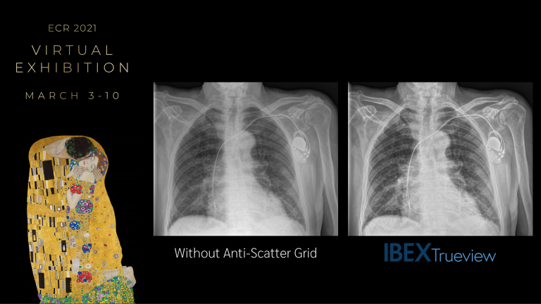
IBEX Trueview® software addresses this challenge by creating gridless images that retain diagnostic image quality in the lung field whilst simultaneously enhancing the visualisation of tubes, lines, and mediastinal structures.
Unlike other image processing techniques, this enables a single image to be used for both diagnosis and the safe placement of lines and tubes, providing the potential for improved safety and workflow efficiency.
IBEX Trueview® delivers enhanced visualisation of mediastinal structures, tubes, and lines whilst retaining diagnostic image quality in the lung field.
IBEX Trueview® uses advanced physics modelling and AI to generate highly accurate scatter estimates and additional compositional outputs from standard radiographs. This is used to remove the effect of scatter and guide advanced post-processing methods with a local knowledge of scatter and composition.
The result: The highest possible diagnostic quality in the lung field with a simultaneous boost in the contrast and visibility of lines, tubes, and bone structures in a single image.
In a recent scoring trial of mobile chest X-ray scans in an ITU setting, clinicians were 3.83⁽¹⁾ times more likely to agree that safe placement of lines and tubes could be confirmed with Trueview images when compared with the original manufacturer images.
(1) 95% confidence interval [2.54,5.83]
Trueview delivers enhanced visualisation of mediastinal structures, tubes, and lines whilst retaining diagnostic image quality in the lung field
Find out more
Head over to our website to learn more about how Trueview can generate higher quality images and new diagnostic outputs including a measure of bone health from standard radiographs.